CELL
TRANSPORT VIDEOS
(Supports 4th Edition, Release date summer of 2021)
|
|
CELL
TRANSPORT VIDEOS |
|
|
Page Topics Elodea Osmosis, Elodea Hypertonic, Elodea Hypotonic, Blood Isotonic, Blood Hypertonic, Blood Hypotonic, Paramecium Contractile Vacuole, Bulk Transport, Paramecium Endocytosis, Paramecium Exocytosis. Brownian movement (motion) is the random movement of suspended particles in a gas or liquid that is caused by the molecular motion (collisions) of surrounding molecules (and particles) of the medium. Two primary factors that influence the rate of movement are temperature and the size of the suspended particles. The rate of movement increases with temperature, and smaller particles move faster because they have less mass (thus, less resistance). The following videos demonstrate the Brownian motion of particles in milk and in india ink. Milk contains an assortment of different sized particles (mostly fat globules), and india ink contains an assortment of different sized carbon particles. WATCH VIDEO OF BROWNIAN MOTION IN INDIA INK WATCH VIDEO OF BROWNIAN MOTION IN MILK In biological systems osmosis is defined as the diffusion of water (from an area of high concentration to an area of low concentration) across a selectively permeable membrane. The concentrations of water vary because of the presence/absence of non-diffusible solute(s). Solutes that are permeable to the membrane reach equilibrium on each side of the selectively permeable membrane. In the following illustration, the permeable solutes, Na+ and Cl- reach equilibrium because they are permeable to the membrane. In the following illustration "A" and "B" start with the same volume of solutions. However, water will move to side "A" because the solution of side "A" has less water (more fructose). The movement of water is an example of osmosis, and as the volume of water in side "A" increases so does its hydrostatic pressure. The separation of the solutes (fructose stays separated from side "B") is an example of dialysis.
Elodea is a common freshwater plant that is frequently used to decorate aquariums. The leaves of Elodea are only two cells thick and are ideal for the microscopic study of the effects of osmotic solutions. Freshwater is hypotonic to Elodea and maintains normal turgor (osmotic) pressure in the plant. A hypotonic solution is a solution that contains less (hypo) solutes than the cytoplasm of the cell. Thus, a hypotonic solution has more water than the cell and water has a tendency to move (diffuse) into the cell. In plants this inward "pressure" produces rigidity of the cell as the plasma membranes are pushed against the cell walls. Plant cells do not rupture because the cell walls resist the outward expansion of the plasma membrane.
WATCH VIDEO OF FRESH (NORMAL) ELODEA A hypertonic solution is a solution that contains more (hyper) solutes than the cytoplasm of the cell. Thus, a hypertonic solution has less water than the cell and water moves (diffuses) out of the cell. As water moves out of the cells there is a loss of turgor pressure and the plasma membranes detach from the cell walls as the cells shrink.
WATCH VIDEO OF NORMAL ELODEA SUBJECTED TO A HYPERTONIC SOLUTION Elodea - Hypertonic Cells in a Hypotonic Solution A hypotonic solution contains less solute (thus, more water) than the cytoplasm of the cells. Thus, placing crenated Elodea cells into a hypotonic solution (100% water) causes water movement into of the cells resulting in the swelling of the cells. The movement of water into the plasmolyzed cells results in an increase in turgor pressure and the cells expand and force the plasma membrane against the cell walls. The cells will not rupture because their turgor pressure is not strong enough to break the walls.
WATCH VIDEO OF PLASMOLYZED ELODEA SUBJECTED TO A HYPOTONIC SOLUTION Normal red blood cells (RBCs, or erythrocytes) are anucleate and appear as small biconcave discs. They contain mostly hemoglobin which gives them a reddish coloration. The plasma membranes of RBCs are extremely fragile and makes the cells ideal for the study of the effects of osmosis. Handling of unscreened human blood in the laboratory requires strict safety requirements and is not recommended. Solutions that contain the same concentration of solutes as the cytoplasm are called isotonic (or normal saline) solutions. Since the solution surrounding the cells and the cytoplasm contain the same concentration of solutes, they contain the same concentrations of water, and there is no net movement of water either into or out of the cell. Thus, the cells maintain their normal shape.
WATCH VIDEO OF BLOOD IN AN ISOTONIC SOLUTION A hypertonic solution contains more solute (thus, less water) than the cytoplasm of the cells. Thus, placing normal RBCs into a hypertonic solution causes water movement out of the cells resulting in crenation. Crenated RBCs are characterized by having scalloped (ridged) membranes. When a RBC loses water, its plasma membrane folds upon itself resulting in a "wrinkled," "scalloped," or "shriveled" appearance called crenated.
WATCH VIDEO OF NORMAL RBCs SUBJECTED TO A HYPERTONIC SOLUTION A hypotonic solution contains less solute (thus, more water) than the cytoplasm of the cells. Thus, placing normal RBCs into a hypotonic solution (100% water) causes water movement into of the cells resulting in the swelling and lysis of the cells. As the RBCs swell the increased turgor (pressure) results in small tears (lysis) forming in the plasma membranes. The tears allow the hemoglobin to begin washing out of the cells resulting in a loss of density and color, or "ghost cells." The cells quickly become totally transparent producing a red colored transparent solution of hemoglobin, plasma membranes, and water. For a more dramatic effect, instead of using normal RBCs the following photographs and video show crenated cells exposed to a hypotonic solution.
WATCH VIDEO OF CRENATED RBCs SUBJECTED TO A HYPOTONIC SOLUTION The cells of the outer layer of the red onion's leaves (bulb scales) are pigmented red. This pigmentation makes the cells easy to identify and study when subjected to solutions that produce the osmotic effects seen in the following video. WATCH VIDEO OF OSMOSIS IN RED ONION Paramecium - Contractile Vacuoles and Osmosis Paramecium is a freshwater protozoa (single-cell eukaryotes) and may be obtained by culture (purchased or maintained in the laboratory) or in the field by sampling scum of stagnant water. Freshwater is hypotonic to Paramecium, and results in the osmotic movement of water into the cell. Specialized organelles called contractile vacuoles function in the homeostatic maintenance of normal turgor pressure (water volume), or osmoregulation. A contractile vacuole is surrounded by radial canals which absorb water from the cytoplasm and move it into the contractile vacuole. Once filled with water, the contractile vacuole contracts and forces the water into the extracellular environment. Upon relaxation, the vacuole refills and then repeats its pumping activity.
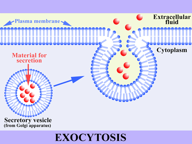
WATCH VIDEO OF CONTRACTILE VACUOLES OF PARAMECIUM Bulk transport (vesicular transport) is the term given to the movement of substances into and out of the cell by membranous organelles called vesicles (vesicular transport). Bulk transport includes endocytosis and exocytosis.
Formation of Food Vacuoles Formation of food vacuoles is an example of endocytosis. Food substances such as bacteria and other protozoa are moved down the oral groove (gullet) and captured by invagination of the plasma membrane in the formation of food vacuoles. The following illustrations and video show Paramecium feeding on yeast that are stained with eosin. Notice that some of the food vacuoles contain yeast that are blue. The blue coloration results from eosin's color change due to a change in the pH of the food vacuoles. The change in pH results as a result of digestive enzymes that are secreted into the food vacuoles.
WATCH VIDEO OF ENDOCYTOSIS, FORMATION OF FOOD VACUOLES OF PARAMECIUM Waste substances are removed from the Paramecium by excretion, an example of exocytosis. Food vacuoles with undesirable or undigested substances are moved to and fused with the plasma membrane resulting in the release of their contents to the extracellular environment. The following illustrations and video show Paramecium in the process of exocytosis.
WATCH VIDEO OF EXOCYTOSIS, SECRETION OF FOOD VACUOLES FROM PARAMECIUM | |