CELL VIDEOS
Supports 4th Edition, Release Date Summer 2021,
|
|
CELL VIDEOS Supports 4th Edition, Release Date Summer 2021, |
|
|
Page Topics Paramecium, Amoeba (Ameba), Euglena, Rotifer (Cilia), Flagellum (Sperm), Cilia (Clam's Gill), Protozoa (Termite's Gut), Adipose Tissue, Ciliated Columnar Epithelium, Blood, Smooth Muscle, Sperm Cells, Stratified Squamous Epithelium, Exfoliated Squamous Epithelium, Links to Cell Videos and Animations. Paramecium is a genus of the ciliate protozoa (Kingdom Protista). Protozoa are the group of single-cell eukaryotes (organisms with nuclei) that usually show mobility and heterotrophy. Heterotrophic organisms derive their carbon from organic compounds and include all animals, fungi, and many bacteria. Autotrophic organisms produce organic compounds by using carbon dioxide as their only carbon source and include plants and some bacteria. Paramecium is a freshwater protozoa and may be obtained by culture (purchased or maintained in the laboratory) or in the field by sampling scum of stagnant water. Paramecium is an ideal laboratory organism for studying the general features of animal cells because they are large and show many characteristics of animal cells. The following features are emphasized on the video: cilia, oral groove, food vacuoles, and contractile vacuoles. Also, notice the flexibility of the plasma membrane and the organism's ability to respond to its environment by changing direction, etc.
CILIA Watch Video of Cilia and other Organelles
ORAL GROOVE
FOOD VACUOLES Watch Video of Food Vacuoles - Endocytosis at Oral Groove
CONTRACTILE VACUOLES Watch Video of Contractile Vacuoles (Osmotic Regulation)
Amoeba is a genus of the protozoa (Kingdom Protista) and move by the formation of temporary "false-feet" called pseudopodia. Amoeba live in a variety of habitats including freshwater, oceans, soil, and as parasites on and within many animals. A common freshwater Amoeba is easily cultured and supplied for laboratory study. Amoeba engulf their prey or food substances by extending pseudopodia around it and capturing it in newly formed cytoplasmic vacuoles, a process called phagocytosis (endocytosis). The video shows pseudopods and the beginning of phagocytosis (endocytosis). (Phagocytosis is also used by many of the body's protective cells, the white blood cells (leukocytes), to engulf and destroy foreign organisms or substances.) Digestive enzymes enter the vacuoles where digestion occurs. The products of digestion move into the cell where they are used in cellular metabolism. Unusable substances are transported by the vacuole to the plasma membrane where they are released into the extracellular environment (egestion, or exocytosis). Amoeba (except marine amoeba) also contain contractile vacuoles that function in osmoregulation. However, the radiating canals associated with the contractile vacuole of Paramecium are not seen.
Euglena is a genus of the flagellate protozoa (Kingdom Protista). Euglena is commonly found in freshwater which is rich in organic nutrients and is easily cultured for laboratory study. Most Euglena are long cylindrical-to-oval in shape and have a single long flagellum that is used for mobility. (The sperm is the only flagellated cell of the human.) Most Euglena contain numerous green chloroplasts and a photo-sensitive structure called the eyespot. Euglena are a favorite food for Amoeba, which captures them by the process of phagocytosis. Newly captured Euglena may be observed in vacuoles within the cytoplasm of Amoeba.
Rotifers are a phylum of mostly freshwater microscopic animals which typically have a pair of wheel-like ciliated tuffs surrounding their mouth. The ciliated "wheels" produce a water current that moves food substances into their mouth where in the pharynx the food is chewed by "jaws." Notice the ciliated tuffs and the "jaws" on the following video. Rotifers usually attach to a surface by a pair of tiny toe-like structures. When unattached the ciliated "wheels" supply the power to move the organism through the water.
Sperm provide the mechanism for the transport of the male's chromosomes. A sperm cell consists of three regions, a head, a mid-piece, and a tail (flagellum). The head mostly contains the nucleus, which contains 23 chromosomes (one of each homologous pair of the diploid cell). The mid-piece mostly contains mitochondria, which supply the energy (ATP) to drive the movement of the tail, the single flagellum.
The gills of clams are lined with ciliated epithelium which functions in producing water currents and the movement of mucus. As water moves into the shell of the clam, its food substances are trapped by the mucus which covers the gills. This video shows the cilia and mucus on the surface of the gill of a clam. Ciliated columnar epithelium is found in the human, especially in the airways of the respiratory system. In the airways of the lungs, the cilia provide for the movement of mucus, which functions to trap particulate substances in inhaled air.
CILIA - INCLUDES VIDEO OF CLAM'S GILL Protozoa in Termite's Intestine (Cilia) Termites eat cellulose, a complex polysaccharide (carbohydrate) of plants. However, termites must first depend upon symbiotic protozoa and bacteria to digest the cellulose to a form which can be absorbed across the termite's gut. This video shows several of the symbiotic protozoa and bacteria in a preparation of the gut of a termite. Notice the numerous cilia on the surface of the protozoa. In the human, the large intestine contains the intestinal (enteric) bacteria, or gut flora. The bacteria (gut flora) function in a symbiotic relationship where the body supplies a nutrient rich environment and the bacteria produce some vitamins and chemically process the intestinal contents.
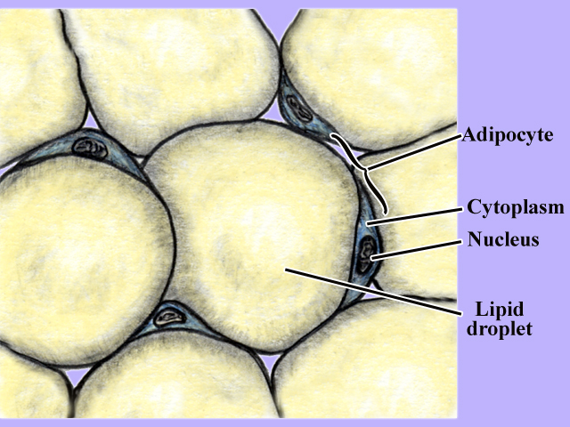
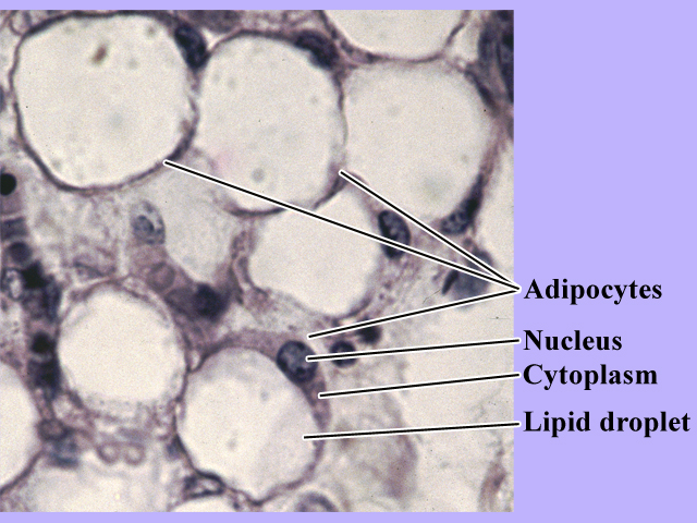
WATCH VIDEO OF TERMITE'S PROTOZOA Adipose tissue functions in lipid (triglyceride) metabolism. Distributed throughout the body mature adipose tissue consists of cells that contain a large lipid droplet of triglycerides. In the well fed state, lipids are stored in adipocytes as a reserve energy source. In times of reduced food consumption (or increased energy demands), the triglycerides are catabolized and mostly used as a source of energy.
WATCH VIDEO SHOWING ADIPOSE TISSUE Ciliated columnar epithelium is a membranous covering tissue (epithelium) that functions in the movement of substances over its surface. Locations include the lining of respiratory airways (such as the trachea, bronchi, and bronchioles) and the fallopian tubes. The following illustrations and video show the ciliated columnar epithelium of the trachea, where the tissue functions in the movement of mucus.
WATCH VIDEO SHOWING CILIATED COLUMNAR EPITHELIUM Blood consists of two major components, a liquid portion (1) the plasma, and the (2) formed elements. The formed elements include the (1) cellular components, the red blood cells (erythrocytes) and the white blood cells (leukocytes) and the (2) non-cellular fragments, the platelets. A blood smear preparation stained with a differential stain (contains several dyes) reveals the different formed elements. The red blood cells do not contain a nucleus (anucleate) and contain mostly hemoglobin. The red blood cells out number the white blood cells by about 800 to 1 and are usually stained a pale shade of red. All of the white blood cells are nucleated and some have distinctively stained cytoplasmic granules. The nuclei of the white blood cells are stained a shade of blue-to-purple, with the granules, if present, stained either blue or red. The white blood cells include the neutrophils, basophils, eosinophils, lymphocytes, and monocytes.
WATCH VIDEO SHOWING BLOOD CELLS WATCH VIDEO SHOWING FRESH BLOOD FROM FINGER STICK Smooth muscle is commonly found in the walls of the body's internal hollow organs, especially those of the digestive, reproductive, and respiratory systems. Under control of the autonomic nervous system, smooth muscle functions in the regulation of the movements of materials in the organs it surrounds. The following illustrations and video show the smooth muscle found in the circular muscle layer of the intestines.
WATCH VIDEO SHOWING SMOOTH MUSCLE CELLS Sperm provide the mechanism for the transport of the male's chromosomes. A sperm cell consists of three regions, a head, a mid-piece, and a tail (flagellum). The head mostly contains the nucleus, which contains 23 chromosomes (one of each homologous pair of the diploid cell). The mid-piece mostly contains mitochondria, which supply the energy (ATP) to drive the movement of the tail, the single flagellum. The following illustrations are from a prepared slide of human sperm. Living sperm are bovine sample.
WATCH VIDEO SHOWING SPERM CELLS PREPARATION WATCH VIDEO SHOWING LIVE BOVINE SPERM CELLS Stratified Squamous Epithelium Stratified squamous epithelium is a membranous covering tissue (epithelium) that functions in protection. Two major regions of the tissue include the basal layer and the surface layer. The basal layer consists of cells that rapidly undergo mitotic divisions, with the newly formed cells being pushed toward the surface. At the surface the cells form the protective surface layer where the old and damaged cells are sloughed. Locations include the lining of mouth, esophagus, vagina, and skin. The following illustrations and video show the stratified squamous epithelium of the esophagus.
WATCH VIDEO SHOWING STRATIFIED SQUAMOUS EPITHELIUM The surface cells of stratified squamous epithelium are easily removed by physical abrasion. The following illustrations and video show stained exfoliated cells obtained by gently scraping the oral mucosa (inner layer of cheek) with a toothpick.
WATCH VIDEO SHOWING STRATIFIED SQUAMOUS EPITHELIUM WATCH VIDEO SHOWING EXFOLIATED SQUAMOUS CELLS LINKS to CELL VIDEOS and ANIMATIONS The following external LINKS contain excellent videos. "Nikon - Microscopy U, A Source for Microscopy Education" Olympus Microscopy Resource Center | |